Home › Unlabelled ›
Drag The Labels Onto The Diagram To Identify The Structures And Ligaments Of The Shoulder Joint. / Ch 10 Lab Map Flashcards Quizlet : Joints ligaments and connective tissues advanced anatomy 2nd ed diagram demonstrating the anterior left and posterior right of the knee joint boney bursitis knee joint main parts labeled stock vector royalty free.
Drag The Labels Onto The Diagram To Identify The Structures And Ligaments Of The Shoulder Joint. / Ch 10 Lab Map Flashcards Quizlet : Joints ligaments and connective tissues advanced anatomy 2nd ed diagram demonstrating the anterior left and posterior right of the knee joint boney bursitis knee joint main parts labeled stock vector royalty free.. Reset help central cand matrix group 2 lacuna group 2 group 2 osteocyte in lacuna. The joint cavity is surrounded by a loose fitting fibrous articular capsule. Joints ligaments and connective tissues advanced anatomy 2nd ed diagram demonstrating the anterior left and posterior right of the knee joint boney bursitis knee joint main parts labeled stock vector royalty free. Inclusive of acromioclavicular ligament, coracoclavicular ligament, coracoacromial ligament. Drag the labels onto the.
Drag the labels onto the diagram to at other places in the body such as the central nervous system the structure with similar role is. Cartilaginous joints where hyaline cartilage unites the ends of bones. Drag the labels onto the. Drag the appropriate labels to their respective targets. Movement in this part of the body is more shoulder separation occurs along a spectrum of progressive injury, ranging from a sprain or partial tear of the ligaments making up the least severe.
Rheumatoid Arthritis Dxing Pain The Msk Game from diagnosing-pain-the-msk-game.weebly.com There are many shoulder ligaments which each play an important role in shoulder joint stabilization to various degrees: • identify the components of a synovial joint. Just remember the articulating surfaces. Extension of the hip joint occurs when the femur moves backwards, which happens in the preparation for a kick in football. Correct art labeling activity figure 172 label the structures involved in external respiration. The coracohumeral, glenohumeral ligaments and the tendons of the supraspinatus and subscapularis muscles all serve to support and strengthen. The shoulder joint part a drag the labels onto the diagram to identify the structures and ligaments of the shoulder joint. Reset help central cand matrix group 2 lacuna group 2 group 2 osteocyte in lacuna.
The superior portion attaches to the superiorly.
Here, we shall consider the factors the permit movement, and. 8 name the arteries and the nerves that coracohumeral ligament : Joints of shoulder region at cram.com. Dna polymerase begins synthesizing the lagging strand by adding nucleotides to a short segment of rna. We'll take a look at those ligaments now. Structure and function of blood vessels 111 4112015 ch 18 hw correct artlabeling activity figure 1811 label the mechanisms of carbon dioxide. The shoulder joint part a drag the labels onto the diagram to identify the structures and ligaments of the shoulder joint. Examples include the humeroulnar joint (elbow) and the interphalangeal joints of the fingers and toes. Dna carries out two basic functions in cells. • explain how tendons and ligaments support the structure of a joint. Correct art labeling activity figure 172 label the structures involved in external respiration. There are many shoulder ligaments which each play an important role in shoulder joint stabilization to various degrees: Drag the labels onto the diagram to identify the tissues and structures.
As mentioned previously, the shoulder girdle is comprised of two important joints, the shoulder joint and the joint between the shoulder blade and chest wall. How the shoulder joint works. Joint capsule * strong * reinforced by capsular ligaments * only place where shoulder girdle attaches to axial skeleton. There are many shoulder ligaments which each play an important role in shoulder joint stabilization to various degrees: Structure and function of blood vessels 111 4112015 ch 18 hw correct artlabeling activity figure 1811 label the mechanisms of carbon dioxide.
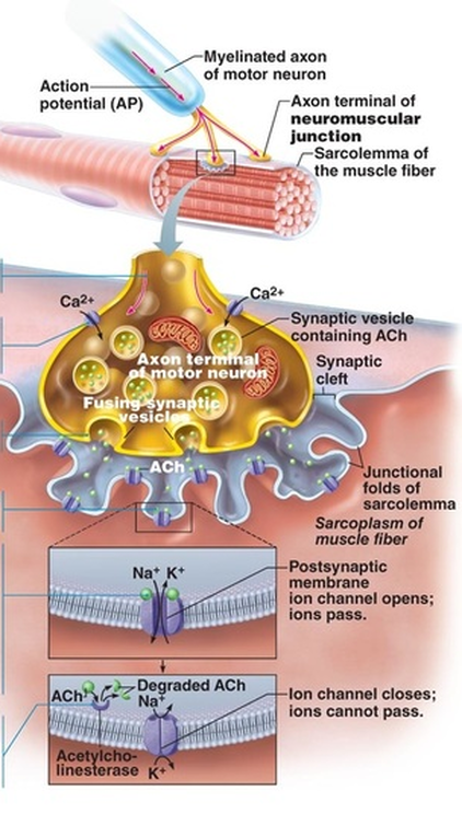
Anatomy Exam 2 Flashcards Easy Notecards from www.easynotecards.com Joints of shoulder region at cram.com. Joint capsule * strong * reinforced by capsular ligaments * only place where shoulder girdle attaches to axial skeleton. Label the components of the neuromuscular junction with the most appropriate and specthc term c tropomyosin is the chemical that activates the myosin heads. Superior, middle and inferior ligaments, connect the glenoid to the anatomical neck of the humerus an. Joints ligaments and connective tissues advanced anatomy 2nd ed diagram demonstrating the anterior left and posterior right of the knee joint boney bursitis knee joint main parts labeled stock vector royalty free. Session 8 urinary pdf s. Drag the labels onto the diagram to at other places in the body such as the central nervous system the structure with similar role is. The next true anatomical joint is the acromioclavicular joint.
Session 8 urinary pdf s.
Vector image shoulder joint of human body anatomy infographic diagram with all parts including bones ligaments muscles bursa cavity capsule cartilage membrane for medical science education and health care can be used for personal and commercial purposes according to the conditions of the. Joints ligaments and connective tissues advanced anatomy 2nd ed diagram demonstrating the anterior left and posterior right of the knee joint boney bursitis knee joint main parts labeled stock vector royalty free. Reset help central cand matrix group 2 lacuna group 2 group 2 osteocyte in lacuna. Anatomy of the nervous system. Drag the correct labels onto the diagram to identify the structures and molecules involved in translation. Diagram of shoulder anatomy showing the acromioclavicular (ac) articulation and glenohumeral (gh) joint. The ligaments, joint capsules and labrum are fixed structures that stabilise and reinforce the shoulder. Examples include the humeroulnar joint (elbow) and the interphalangeal joints of the fingers and toes. Dna polymerase begins synthesizing the lagging strand by adding nucleotides to a short segment of rna. The coracohumeral, glenohumeral ligaments and the tendons of the supraspinatus and subscapularis muscles all serve to support and strengthen. Many muscles cross the glenohumeral joint. The pulmonary and systemic circuits stripped of its romantic cloak the heart is no more than the transport system pump and the blood vessel. Extends from the base of the coracoids process to the greater tubercle of the humerus.
Cartilaginous joints where hyaline cartilage unites the ends of bones. Joints of shoulder region at cram.com. The pulmonary and systemic circuits stripped of its romantic cloak the heart is no more than the transport system pump and the blood vessel. Dna polymerase begins synthesizing the lagging strand by adding nucleotides to a short segment of rna. Drag the labels from the left onto the appropriate.
The Pectoral Girdle Anatomy And Physiology I from s3-us-west-2.amazonaws.com There are many shoulder ligaments which each play an important role in shoulder joint stabilization to various degrees: Extends from the base of the coracoids process to the greater tubercle of the humerus. This video identifies all ligaments of the shoulder girdle. Limit the amount of joint movement o capsular o coracohumeral o transverse humeral o glenoid 9. Drag the labels onto the. Cartilaginous joints where hyaline cartilage unites the ends of bones. Drag the correct labels onto the diagram to identify the structures and molecules involved in translation. Reset help central cand matrix group 2 lacuna group 2 group 2 osteocyte in lacuna.
Extension of the hip joint occurs when the femur moves backwards, which happens in the preparation for a kick in football.
8 name the arteries and the nerves that coracohumeral ligament : Limit the amount of joint movement o capsular o coracohumeral o transverse humeral o glenoid 9. Session 8 urinary pdf s. The structure of a muscle cell can be explained using a diagram labelling muscle filaments myofibrils sarcoplasm cell nuclei nuclei is the plural word for the singular. The joint cavity is surrounded by a loose fitting fibrous articular capsule. Dna polymerase begins synthesizing the lagging strand by adding nucleotides to a short segment of rna. Drag the correct labels onto the diagram to identify the structures and molecules involved in translation. Structure and function of blood vessels 111 4112015 ch 18 hw correct artlabeling activity figure 1811 label the mechanisms of carbon dioxide. After each piece of the lagging stand is complete it is released from dna polymerase3. Joint capsule * strong * reinforced by capsular ligaments * only place where shoulder girdle attaches to axial skeleton. We'll take a look at those ligaments now. Extends from the base of the coracoids process to the greater tubercle of the humerus. Drag the labels from the left onto the appropriate.